
ملف:HIV entry into T cell schematic.png

الملف الأصلي (2٬805 × 3٬405 بكسل حجم الملف: 4٫49 ميجابايت، نوع MIME: image/png)
| هذا ملف من ويكيميديا كومنز. معلومات من صفحة وصفه مبينة في الأسفل. كومنز مستودع ملفات ميديا ذو رخصة حرة. |
ملخص

|
هذه biology الصورة / الصورتان باستعمال رسومات متجهية ملفات رسوميات شعاعية.
It is recommended to name the SVG file "HIV entry into T cell schematic.svg" - then the template Vector version available (or Vva) does not need the new image name parameter.
|
| الوصف |
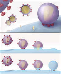
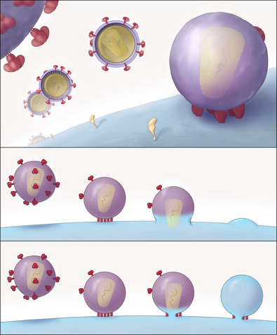
English: "Schematic Representation of the Key Structural Features of SIV and HIV-1 Entry into T Cells"
(A) Different stages of viral entry from budding, to maturation, to entry claw formation. For the SIV strain used here, viruses that are docked to the cell via an entry claw show very few, if any, viral spikes on their surface, whereas non-contacting viruses typically display between 60 and 100 spikes on their surface. The entry claw is composed of between five to seven anchors spanning the region between the virus and the cell, each ∼100 Å long, and spaced laterally by ∼150 Å. (B and C) Two alternative models for viral entry. In the global fusion model (B), the formation of the entry claw is followed by progressive fusion of the viral membrane across its width, leading to merger of the contents of the viral membrane with the cellular membrane. In the local fusion model (C), the formation of the entry claw is followed by the creation of a local pore centered at one of the rods, leading to delivery of the viral core into the cell." |
| التاريخ | Published May 4, 2007 |
| المصدر |
Sougrat R, Bartesaghi A, Lifson JD, et al (May 2007). "Electron tomography of the contact between T cells and SIV/HIV-1: implications for viral entry". PLoS Pathog. 3 (5): e63. PMID 17480119. doi:10.1371/journal.ppat.0030063 Direct link to image: http://www.plospathogens.org/article/showImageLarge.action?uri=info%3Adoi%2F10.1371%2Fjournal.ppat.0030063.g008 |
| المؤلف | Rachid Sougrat, Alberto Bartesaghi, Jeffrey D. Lifson, Adam E. Bennett, Julian W. Bess, Daniel J. Zabransky, Sriram Subramaniam |
| الترخيص (إعادة استخدام هذا الملف) |
[1] |
| إصدارات أخرى | JPG version |
ترخيص
|
هذا الملف مُرخص تحت رخصة المشاع المبدع نسبة المصنف إلى مؤلفه 2.5 العامة
|
نُشِر هذا الملفُّ في دورية المكتبة العامَّة للعلوم. يُصرِّح الموقع الرسميِّ بأنَّ مُحتويات دوريات المكتبة العامَّة للعلوم مَنشورةٌ جميعها تحت رخصة المَشاع الإِبداعيِّ نسبة المُصنَّف إِلى مُؤَلِّفه (أو إلى إصداراتٍ سابِقة مِن الرُّخصة حسب تاريخ الإِصدار)، ما لم يذكر خلاف ذلك. |
تاريخ الملف
اضغط على زمن/تاريخ لرؤية الملف كما بدا في هذا الزمن.
| زمن/تاريخ | صورة مصغرة | الأبعاد | مستخدم | تعليق | |
|---|---|---|---|---|---|
| حالي | 00:30، 11 يونيو 2008 | | 2٬805 × 3٬405 (4٫49 ميجابايت) | Fvasconcellos | {{Information |Description="Schematic Representation of the Key Structural Features of SIV and HIV-1 Entry into T Cells" (A) Different stages of viral entry from budding, to maturation, to entry claw formation. For the SIV strain used here, viruses that |
استخدام الملف
الصفحة التالية تستخدم هذا الملف:
الاستخدام العالمي للملف
الويكيات الأخرى التالية تستخدم هذا الملف:
- الاستخدام في en.wikipedia.org
- الاستخدام في es.wikipedia.org
- الاستخدام في ko.wikipedia.org
- الاستخدام في outreach.wikimedia.org
- الاستخدام في sl.wikipedia.org